The term colonic atresia describes a condition in which a part of the colon has failed to correctly form, and that part is either completely blocked or is altogether missing. Colonic stenosis describes a condition in which a part of the colon is very narrow, resulting in a partial blockage. Other obstructions of the colon that affect newborns include Hirschsprung disease and small left colon syndrome, as well as obturation obstruction, meconium ileus, or meconium plug. These issues are all forms of colonic obstruction; however, they are different from atresia and stenosis and are more completely reviewed in their own articles.
The colon is the rarest site of atresia in the GI tract. It is a congenital anomaly which may be suggested using prenatal ultrasonography and is usually revealed in affected newborns shortly after birth. Patients usually present with abdominal distention and failure to pass meconium.
Stenosis of the colon is much more common. Patients usually present later in life, most often because of an identifiable event. In congenital stenosis, a narrow segment of colon is observed, but bowel continuity is maintained. A discrepancy between the colonic segments above and below the area of stenosis is present. In acquired stenosis, which is commonly referred to as stricture, what starts as a normal segment becomes narrowed. This is most common in premature babies who have recovered from an episode of necrotizing enterocolitis.
History of the Procedure
Colonic atresia was first recorded in 1673.1 The first survivor was reported by Gaub in 1922 and was treated using a diverting colostomy.2 Potts reported the first survival following primary repair in 1947.3
Problem
The common problem in both atresia and stenosis is intestinal blockage, either partial or complete.
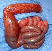
In colonic atresia, the problem is complete bowel obstruction. Gas and stool cannot pass, and the colonic segment above the atresia becomes distended. If left untreated, this leads to perforation.
In colonic stenosis, the problem is that gas and stool try to pass through a narrow area. While the baby is passing soft baby stools, this may or may not be noticeable. When the baby's diet changes from breast milk or formula to cereals and solid foods, the stool can become thicker and more formed. This may cause stenosis to become symptomatic, leading to distension, feeding intolerance, or failure to thrive.
In babies who have had necrotizing enterocolitis, stenosis occurs after the original episode has resolved. This may manifest in varying degrees, ranging from minor feeding intolerance and distension to near-complete bowel obstruction.
Frequency
Colon atresia is very rare. Various incidences have been reported, ranging from 1 in 1500 live births4 to 1 in 66,000 live births.5 Webb (1931) and Benson (1968) cited the incidence as 1 in 20,000, which is most reflective of experiences in the modern era.6,7 In a 1982 report, Powell suggested that colonic atresia represents 5-15% of intestinal atresias,8 whereas in 1966, Freeman reported a lower figure of 1.8%.9 In 1953, Gross recorded 6 cases of colonic atresia out of 140 cases of intestinal atresia (4.3%) at the Boston Children's Hospital.10
Multiple atresias are uncommon in the colon; however, colonic atresia may be overlooked when small intestinal atresia is present.11 Rare cases of familial colonic atresia have been described. Animal studies have shown an autosomal recessive pattern of inheritance in cattle. Hereditary multiple intestinal atresia affects both the large and small intestine, whereas nonhereditary multiple intestinal atresia usually spares the colon.12
The incidence of colonic stenosis is not readily available because most cases are acquired. In 1953, Gross noted a single colonic lesion in 71 patients with intestinal stenosis.10 Necrotizing enterocolitis is the most common etiology of postnatal colonic stenosis; narrowing develops in 10-25% of affected patients.13,14,15
Etiology
See Relevant Anatomy.
Pathophysiology
See Relevant Anatomy.
Presentation
Patients with colonic atresia or congenital stenosis may sometimes have findings on prenatal ultrasonography, such as dilated bowel loops or the presence of polyhydramnios. Initial physical examination findings are normal in the absence of associated conditions. The anus usually appears normal. Progressive abdominal distention develops. Rectal examination reveals white or pale mucus rather than pigmented meconium. Failure to completely pass meconium suggests atresia, whereas delayed passage of meconium (>24 h) suggests Hirschsprung disease. Patients with colonic atresia may pass meconium normally because the incident that caused the atresia may have occurred after the colon had become filled with meconium.
Colonic stenosis usually follows some form of injury to the colon, and ischemia is considered central to the insult. This injury may be in utero or postnatal. The infant or child may present with symptoms similar to atresia with high-grade stenosis; less stenotic lesions may not become apparent until feeding is undertaken. In those instances, the child's abdomen may become distended with feeding, and stool production is scant, if present.
Babies with necrotizing enterocolitis may show signs of acquired stenosis following their acute episode. When the septic signs of the illness resolve and the child is doing well, feeding is often attempted. Those babies who have formed stenoses usually do not tolerate feeds and become distended. Studies may be performed to confirm the diagnosis and to try to localize the site of narrowing, at the discretion of the surgeon.
Associated conditions
Colonic atresia has been associated with abdominal wall defects and abnormalities of the genitourinary tract.16 Nonfixation of the colon has been reported.17 Association with anal atresia18 and imperforate anus19 has been reported but is extremely rare. Colonic perforation may occur.20 This is thought to be caused by overdistension of closed colonic loop, with gas and stool trapped between a competent ileocecal valve proximally and the blind-ending colon distally. In 1988, Pohlson et al reported perforation of the terminal ileum in one case, clearly demonstrating that perforation can occur anywhere proximal to the obstructed bowel.21
Hirschsprung disease has been present in a small number of cases.22,23,24,25 Additional anomalies associated with this pairing include omphalocele26 and absence of a hand.27 In most cases the aganglionosis is discovered after colostomy closure when the distal bowel does not properly function. Some authors have recommended that rectal biopsy be performed at the time of laparotomy,28 whereas others believe that biopsy should be reserved for children who do not pass stool readily upon restoring bowel continuity after resection.23,25 Because colonic atresia is rare and because of the morbidity and mortality associated with missing the diagnosis prior to establishing intestinal continuity, rectal biopsy prior to definitive repair would seem prudent.29
Cardiac conditions that require catheterization may predispose a baby to a mesenteric vascular incident resulting in colonic stenosis. Additional conditions reported in patients with colonic stenosis include cryptophthalmia syndrome (ie, cleft lip and palate, microphthalmia, dysplastic kidneys, proximal jejunal atresia), arthrogryposis, proximal intestinal atresia, neoplasm, and malrotation.4,8 Riley-Day syndrome (ie, familial dysautonomia) has been associated with spontaneous colon ischemia.30 Coloboma, cataracts and facial hemihypertrophy, facial asymmetry with palsy, microphthalmia with partial iridial coloboma, exophthalmia, and bilateral optic nerve hypoplasia all have been reported.8 Kim et al (2000) described a case of colonic atresia in monozygotic twins.12
Indications
Like all other intestinal atresias, colonic atresia is fatal if the obstruction is not relieved. A baby with colonic atresia is at risk for dehydration, perforation, and sepsis. Any intestinal atresia requires operative intervention to prevent these complications.
The specific operative indication in colonic atresia is complete bowel obstruction. Once a newborn is identified as having bowel obstruction, no additional information is required to prove the need for surgery. Other studies may be done prior to operation, but no studies are necessary to confirm the need for surgery.
Colonic stenosis behaves like colonic atresia when the lesion is very tight. In less severe cases, the child may have chronic problems like bloating with feeds, cramping, or poor weight gain. Any of these symptoms may be significant enough to warrant either radiological investigations or operative exploration and repair.
Relevant Anatomy
Embryology, anatomy, and pathophysiologyThe colon arises from the digestive tube, which is present by the end of the first month of gestation. Rapid elongation begins at 5 weeks' gestation. Over the ensuing 5 weeks, the intestinal tube, separable into cephalad and caudal limbs (based on the relationship to the omphalomesenteric duct), rotates counterclockwise and returns to its familiar position in the abdomen. The proximal caudal limb receives its blood supply from the superior mesenteric artery (SMA), whereas the inferior mesenteric artery (IMA) supplies the distal portion.31
The SMA gives rise to the ileocolic, right colic, and middle colic arteries, which supply the ileocecal region, the ascending colon, and the proximal transverse colon, respectively. The left colic artery, arising from the IMA, supplies the left portion of the transverse colon and the descending colon. The middle colic and left colic arteries join near the splenic flexure to form the marginal artery. The sigmoid arteries, rectosigmoid arteries, and branches of the IMA supply the sigmoid colon.
Small bowel and colonic atresias are not believed to occur by the same process as in duodenal atresia, which is suspected to be due to failure of vacuolization of the duodenum and was described by Tandler in 1900.
In 1955, Louw and Barnard hypothesized that small bowel atresias are caused by prenatal vascular interruption.32 The same mechanism is believed to cause colonic atresia. Thrombosis, volvulus, and herniation with strangulation are all mechanisms that may cause in utero vascular injury and bowel necrosis with subsequent reabsorption. Fairbanks proposed a relationship between fibroblast growth factor 10 expression, vascular development, and colonic atresia.33
Although the colon receives its blood supply from the SMA and the IMA, much variation among smaller named arterial branches is observed. The portions of the colon most vulnerable to ischemia appear to be the splenic flexure and the ileocecal area. Coincidentally, these areas are the farthest from the major trunks. Nevertheless, atresia and stenosis occur throughout the colon; the wide variation in terminal branches coupled with the complex rotation and fixation of the bowel may create regions of bowel ischemic injury in utero that result in atretic or stenotic lesions.34
Any process that leads to occlusion of branches of these vessels in utero may result in atresia. Compression at the umbilical ring,35 internal hernia, intussusception,36 choledochal cyst,37 volvulus, and thrombosis have all been implicated in the initiation of bowel infarction, leading to disintegration, gradual reabsorption of dead tissue, and sealing of the bowel ends in the fetus as described by Louw in 1964.38 The bowel content is sterile; thus, sepsis does not occur. Meconium is produced throughout the gut during the third trimester; thus, a newborn whose atresia occurs after that time may still pass meconium in the newborn period.
Prenatal maternal use of vasoconstrictive medications, such as cocaine, amphetamines, nicotine, or decongestants, has been suggested to be a risk factor for intestinal atresia formation.39
Congenital stenosis occurs when the bowel injury is incomplete. This may happen when injury occurs close to the bowel wall, allowing collateral blood flow to preserve the injured tissue. Another mechanism is limited ischemia, in which the blood supply is partially or intermittently occluded, resulting in incomplete intestinal injury.
Acquired stenosis is more common than atresia or congenital stenosis. Via the same mechanism of vascular compromise, the injured bowel undergoes healing and scarring with narrowing of the affected intestine. Intense inflammatory reactions, such as those that occur in necrotizing enterocolitis and Crohn disease, may result in stricture. Tuberculosis-associated left colon stricture has also been reported.40
Patients who undergo bowel resection for any reason and have a segment of intestine removed and joined by anastomosis may develop stricture at the anastomotic site due to ischemia or technical issue. Surgery is usually required to revise the joined loops.
Colon atresia is typically classified using the 1989 descriptions of intestinal atresia by Bland-Sutton and the 1964 descriptions by Louw.41,38 In type 1 lesions, the bowel and mesentery remain intact, but the bowel lumen is interrupted by a complete membrane (see Media file 1). Type 2 lesions are those in which the bowel is discontinuous, connected by a fibrous cord. In type 3 lesions, the bowel ends are completely separated, and the mesentery has a gap. Stenotic lesions are characterized by intact bowel with incomplete occlusion and require no classification.
In 1990, Davenport et al reviewed 118 cases of colonic atresia. The distribution of the lesions by site was as follows:5
- Ascending colon - 33 (28%)
- Hepatic flexure - 4 (3%)
- Transverse colon - 27 (23%)
- Splenic flexure - 30 (25%)
- Descending and sigmoid - 24 (20%)
Two thirds of colon atresias are in the distribution of the IMA. This may relate to lack of collateral blood supply or disease processes that render this portion of the colon more susceptible to injury.
Contraindications
Intestinal obstruction at any level requires surgical relief. The timing of surgery in colonic atresia and stenosis depends on the patient's clinical condition and their associated malformations and comorbidities.
The decision to proceed with primary correction (resection with anastomosis) or stoma diversion depends on the same factors, as well as the skill and experience of the surgeon and their team. Although no contraindications to surgery are recognized, stoma diversion is the minimum intervention necessary to relieve the obstruction. Severe underlying illness and associated life-threatening malformations may be considered relative contraindications to immediate primary repair.